Introduction
Magnetic resonance imaging (MRI) is one of the most advanced medical technologies available today. It provides detailed images of the internal structures of the body using a combination of powerful magnets and radio waves. But how does an MRI scanner actually work? In this article, we’ll explore the anatomy of an MRI machine, the physics behind it, and the step-by-step process of the MRI scan.
What is an MRI Machine?
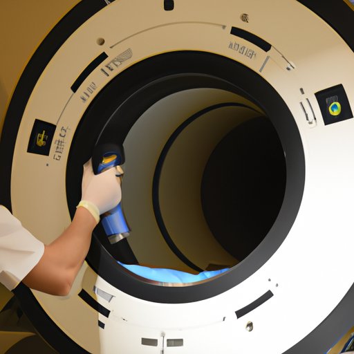
An MRI machine is a large, tube-shaped scanner that uses a powerful magnet and radio waves to create detailed images of the inside of the body. The patient lies on a table that slides into the center of the machine. The MRI scanner is composed of several components, including a powerful magnet, a computer, and a radio frequency coil.

Overview of the Anatomy of an MRI Scanner
The main component of an MRI scanner is the magnet. The magnet creates a strong, uniform magnetic field in the area around the patient. This magnetic field interacts with the body’s hydrogen atoms, causing them to spin in a certain direction. A radio frequency coil is then used to send and receive radio signals from the spinning atoms. The computer then interprets these signals and creates a detailed image of the body’s internal structures.

The Physics Behind Magnetic Resonance Imaging
The physics behind MRI is complex, but it can be explained in simple terms. At the core of MRI is the concept of resonance – when two objects vibrate at the same frequency, they create a powerful force. In the case of MRI, the magnetic field created by the magnet interacts with the body’s hydrogen atoms, causing them to spin in a certain direction. When the radio frequency coil sends out a signal of the same frequency, the spinning atoms resonate with the signal and emit their own signal. This signal is then picked up by the coil and interpreted by the computer, which creates an image of the body’s internal structures.
An Overview of the MRI Procedure
An MRI scan is a non-invasive procedure that typically takes between 30 minutes and an hour. Before the scan begins, the patient is given a set of instructions, such as removing any metal objects or jewelry and avoiding movement during the scan. Depending on the type of scan being performed, the patient may also need to drink a special contrast solution prior to the scan.
Types of MRI Scans
There are several different types of MRI scans that can be used to diagnose different medical conditions. Common types of MRI scans include brain scans, abdominal scans, chest scans, and joint scans. Each type of scan uses a different set of parameters to produce a detailed image of the area being scanned.

Preparation for the MRI Scan
Before the scan begins, the patient is asked to remove all metal objects and jewelry. They are also asked to change into a hospital gown or other clothing without metal zippers or buttons. If a contrast solution is needed, the patient will be instructed to drink it prior to the scan. After this preparation is complete, the patient is ready to begin the scan.
Safety Considerations
MRI scans are generally very safe, but there are some important safety considerations. Patients should avoid moving during the scan, as sudden movements can cause blurry images. Additionally, patients with pacemakers or other metal implants must inform their doctor before the scan, as the powerful magnetic field can interfere with the device. Finally, pregnant women should not undergo an MRI scan unless absolutely necessary, as the effects of the radiation on the fetus are unknown.
A Step-by-Step Look at the MRI Process
Once the patient is prepared, the MRI scan can begin. Here is a step-by-step look at the MRI process:
Prepping the Patient
The patient is asked to lie down on the table and make themselves comfortable. Once the patient is in position, the technician will adjust the table and straps to ensure the patient is secure and won’t move during the scan.
Positioning the Patient
The patient is then positioned within the scanner. The technician will move the table until the desired area is in the center of the scanner. The technician will then use a laser pointer to indicate the exact area to be scanned.
Gathering and Analyzing the Data
Once the patient is in position, the technician will start the scan. During the scan, the computer will gather data from the spinning atoms and construct a detailed image of the area being scanned. This image is then analyzed by the technician and any abnormalities are noted.
Post-Scan Procedures
Once the scan is complete, the patient can leave the scanner and the technician will review the images. The images are then sent to a radiologist, who will interpret the images and provide a diagnosis. Depending on the results of the scan, further tests or treatments may be recommended.
Conclusion
MRI is a powerful medical technology that can provide detailed images of the body’s internal structures. By understanding the anatomy of the MRI machine, the physics behind it, and the step-by-step process of the MRI scan, we can better appreciate the power and potential of this amazing technology. While MRI scans are generally very safe, it’s important to follow all safety instructions and to tell your doctor if you have any metal implants or are pregnant. With proper preparation, an MRI scan can provide invaluable diagnostic information and help doctors make informed decisions about treatment options.
(Note: Is this article not meeting your expectations? Do you have knowledge or insights to share? Unlock new opportunities and expand your reach by joining our authors team. Click Registration to join us and share your expertise with our readers.)
